CT HEAD INTERPRETATION GUIDE
ain ct scan
How do you read a CT brain or ct brain anatomy ?
What does a CT scan of the brain show?
What are they looking for with a CT scan in ct brain ?
Why is CT blood white?
The CT head scan is one of the most common imaging studies that you can be faced with and the most frequently requested by A&E. ct brain interpretation This app will cover some of the underlying principles of CT head studies, and discuss a method for their interpretation. ct scan of brain
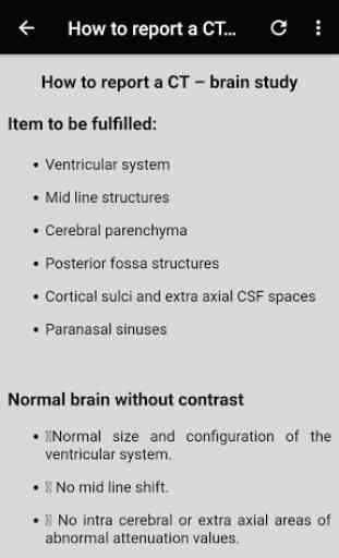
Interpretation of the CT scan of the Brain :
Underlying principles and terminology
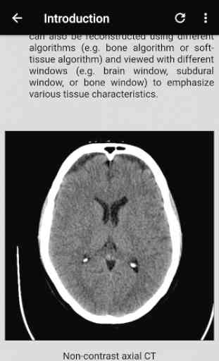
Computed tomography (CT) scanning involves the use of x-rays to take cross-sectional images of the body. This is possible as different tissues interact with X-rays in different ways. Some tissues will allow the passage of these X-rays without influencing it much, whilst other tissues will exert a more significant effect. The extent to which a material can be penetrated by an x-ray beam is described in terms of an attenuation coefficient which assesses how much a beam is weakened by passing through a voxel of tissue (voxel = volumetric pixel).
These values are frequently expressed as Hounsfield Units (HU). Distilled water at standard temperature and pressure has 0 HU, whereas air under the same conditions has -1000 HU. Approximate values for various tissues are outlined in table 1 (these are not set in stone – only rough estimates).
Confirm details ct scan brain anatomy
As with the interpretation of all studies, the first step is to confirm you have the correct patient and scan.
Check the following:
Patient name, hospital number and date of birth
Date and time the scan was acquired
Always look at previous scans so that you have something to compare to
The appearance of tissues on a CT scan is described in terms of ‘density’. Darker structures are ‘hypodense or low density’; brighter structures are ‘hyperdense or high density’.
Blood Can Be Very Bad is a mnemonic that can be used when faced with a CT head scan. Think of this approach as a framework for a quick review of a scan – it won’t turn you into an experienced radiologist! It’s important to recognise that more subtle signs might still be overlooked. Furthermore, you should work through the entire system even if you spot something obvious early on (e.g. if you see a large extradural haematoma, still check the cisterns, brain, ventricles and bone for any other abnormalities).
Blood
Check for evidence of:
Extradural haematoma (Extra-axial)
Subdural haematoma (Extra-axial)
Subarachnoid haemorrhage – may be very subtle. Remember a SAH can extend into the ventricular system so ALWAYS look at the posterior horns as blood may collect in the dependant portion.
Intraventricular haemorrhage
Intraparenchymal haemorrhage (Intra-axial)
Bear in mind that blood will have varying appearances depending on the age of the collection, with a more acute haematoma appearing hyperdense compared to a chronic bleed. Some bleeds may also be very subtle and difficult to spot unless you look closely and this is one of the reasons why windowing is so important.
How do you read a CT brain or ct brain anatomy ?
What does a CT scan of the brain show?
What are they looking for with a CT scan in ct brain ?
Why is CT blood white?
The CT head scan is one of the most common imaging studies that you can be faced with and the most frequently requested by A&E. ct brain interpretation This app will cover some of the underlying principles of CT head studies, and discuss a method for their interpretation. ct scan of brain
Interpretation of the CT scan of the Brain :
Underlying principles and terminology
Computed tomography (CT) scanning involves the use of x-rays to take cross-sectional images of the body. This is possible as different tissues interact with X-rays in different ways. Some tissues will allow the passage of these X-rays without influencing it much, whilst other tissues will exert a more significant effect. The extent to which a material can be penetrated by an x-ray beam is described in terms of an attenuation coefficient which assesses how much a beam is weakened by passing through a voxel of tissue (voxel = volumetric pixel).
These values are frequently expressed as Hounsfield Units (HU). Distilled water at standard temperature and pressure has 0 HU, whereas air under the same conditions has -1000 HU. Approximate values for various tissues are outlined in table 1 (these are not set in stone – only rough estimates).
Confirm details ct scan brain anatomy
As with the interpretation of all studies, the first step is to confirm you have the correct patient and scan.
Check the following:
Patient name, hospital number and date of birth
Date and time the scan was acquired
Always look at previous scans so that you have something to compare to
The appearance of tissues on a CT scan is described in terms of ‘density’. Darker structures are ‘hypodense or low density’; brighter structures are ‘hyperdense or high density’.
Blood Can Be Very Bad is a mnemonic that can be used when faced with a CT head scan. Think of this approach as a framework for a quick review of a scan – it won’t turn you into an experienced radiologist! It’s important to recognise that more subtle signs might still be overlooked. Furthermore, you should work through the entire system even if you spot something obvious early on (e.g. if you see a large extradural haematoma, still check the cisterns, brain, ventricles and bone for any other abnormalities).
Blood
Check for evidence of:
Extradural haematoma (Extra-axial)
Subdural haematoma (Extra-axial)
Subarachnoid haemorrhage – may be very subtle. Remember a SAH can extend into the ventricular system so ALWAYS look at the posterior horns as blood may collect in the dependant portion.
Intraventricular haemorrhage
Intraparenchymal haemorrhage (Intra-axial)
Bear in mind that blood will have varying appearances depending on the age of the collection, with a more acute haematoma appearing hyperdense compared to a chronic bleed. Some bleeds may also be very subtle and difficult to spot unless you look closely and this is one of the reasons why windowing is so important.
Category : Medical
Related searches
Reviews (3)
Ahm. H.
Sep 30, 2019
Best app for the CT brain
Alm. K.
Sep 10, 2019
Best app
best app for head CT basics