Endropoda
Endropoda (Educational Android Apps for Cancer Radiotherapy Planning) is an edugame application that is designed for radiotherapy course supplement, especially for medical physics and medical students. Endropoda was developed by medical physics student of University of Brawijaya collaborating as a realization of student creativity program that was supported by Ministry of Education and Culture, Indonesia.
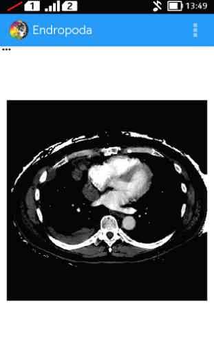
Endropoda is an educational app for external radiotherapy planning which can simulate beam penetration ability based on attenuation characteristic of a medium (patient organ). The dose distribution of radiation source that is placed in a distant from patient skin is displayed as area with same colour (as if basic principles of isodose curve). Endropoda can calculate automatically the absorbed intensity (that is proportional with absorbed dose of an organ), the dose then will be displayed as colour spectrum representing the amount of absorbed dose in the organ. In addition, Endropoda can identify the type of an organ based on radiation attenuation characteristic (CT-Number value). Endropoda is also have an example of 3D reconstruction of CT-Scan image that can help user to imagine the anatomical feature of human body.
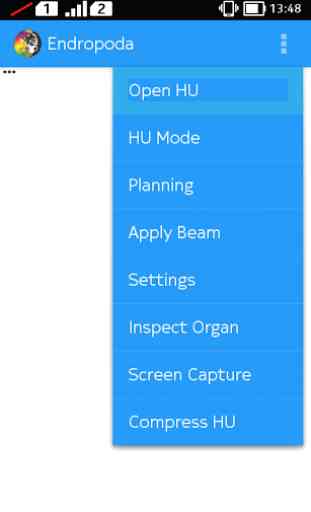
ENDROPODA contains five main features:
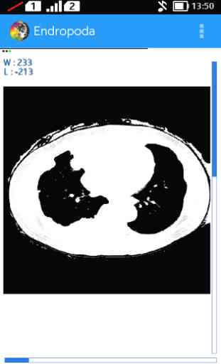
- HU mode : In this mode we can set the window width and window level of CT-Scan image to produce higher contrast higher contrast for certain organ, so it will be displayed better than another organ its surrounding. The basic principal of window technique is based on CT number. Where is CT number is a value related to attenuation coefficient of certain organ that can be used to distinguish an organ from another based on their attenuation coefficient.
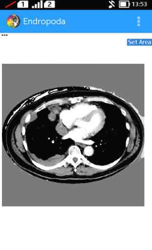
- Planning : In planning mode, user can create a region of interest on CT-Scan image to eliminate the background area and also can create cancer area that will be treated with radiation
- Apply Beam : User can see the Beam direction for cancer treatment, including the colour distribution describes the absorbed dose
- Inspect Organ : The organ identification will be displayed automatically based on CT Number value that is stored in each pixel of CT-Scan image
- En-Link: http://lovinaliu.blogspot.com/ is a Link to all information about external radiotherapy, especially for Cobalt-60 source (in Indonesian). This link also provides an example of 3D reconstruction of CT-Scan image that can help user to imagine the anatomical feature of human body.
A modified CT-Scan file example can be downloaded at:
https://www.dropbox.com/l/gocY1J5peRkVlxQOZGK0jq
Resume of Radiotherapy lecture and tutorial about how to use Endropoda can be found at:
http://lovinaliu.blogspot.com/
DISCLAIMER :
• All of data and information displayed in Endropoda has been through ethical clearance procedure for educational purpose only. It was not designed for medical decision
• External radiotherapy planning procedure that is displayed in Endropoda is intended as basic introduction about external radiotherapy. It does not replace the full length protocol as is used in hospital.
• The displayed isodose curve and dose calculation are based on intensity attenuation. It does not describe beam penetration ability for certain type of radiation.
• CT-Scan data used in Endropoda was generated by the developer and are not taken from patients (it is not contain anything data of real patient)
Future version of ENDROPODA will include additional features based on user feedback.
Contact person :Lovina Wijayanti ([email protected])Ubaidillah ([email protected])
Endropoda is an educational app for external radiotherapy planning which can simulate beam penetration ability based on attenuation characteristic of a medium (patient organ). The dose distribution of radiation source that is placed in a distant from patient skin is displayed as area with same colour (as if basic principles of isodose curve). Endropoda can calculate automatically the absorbed intensity (that is proportional with absorbed dose of an organ), the dose then will be displayed as colour spectrum representing the amount of absorbed dose in the organ. In addition, Endropoda can identify the type of an organ based on radiation attenuation characteristic (CT-Number value). Endropoda is also have an example of 3D reconstruction of CT-Scan image that can help user to imagine the anatomical feature of human body.
ENDROPODA contains five main features:
- HU mode : In this mode we can set the window width and window level of CT-Scan image to produce higher contrast higher contrast for certain organ, so it will be displayed better than another organ its surrounding. The basic principal of window technique is based on CT number. Where is CT number is a value related to attenuation coefficient of certain organ that can be used to distinguish an organ from another based on their attenuation coefficient.
- Planning : In planning mode, user can create a region of interest on CT-Scan image to eliminate the background area and also can create cancer area that will be treated with radiation
- Apply Beam : User can see the Beam direction for cancer treatment, including the colour distribution describes the absorbed dose
- Inspect Organ : The organ identification will be displayed automatically based on CT Number value that is stored in each pixel of CT-Scan image
- En-Link: http://lovinaliu.blogspot.com/ is a Link to all information about external radiotherapy, especially for Cobalt-60 source (in Indonesian). This link also provides an example of 3D reconstruction of CT-Scan image that can help user to imagine the anatomical feature of human body.
A modified CT-Scan file example can be downloaded at:
https://www.dropbox.com/l/gocY1J5peRkVlxQOZGK0jq
Resume of Radiotherapy lecture and tutorial about how to use Endropoda can be found at:
http://lovinaliu.blogspot.com/
DISCLAIMER :
• All of data and information displayed in Endropoda has been through ethical clearance procedure for educational purpose only. It was not designed for medical decision
• External radiotherapy planning procedure that is displayed in Endropoda is intended as basic introduction about external radiotherapy. It does not replace the full length protocol as is used in hospital.
• The displayed isodose curve and dose calculation are based on intensity attenuation. It does not describe beam penetration ability for certain type of radiation.
• CT-Scan data used in Endropoda was generated by the developer and are not taken from patients (it is not contain anything data of real patient)
Future version of ENDROPODA will include additional features based on user feedback.
Contact person :Lovina Wijayanti ([email protected])Ubaidillah ([email protected])
Category : Education
Related searches