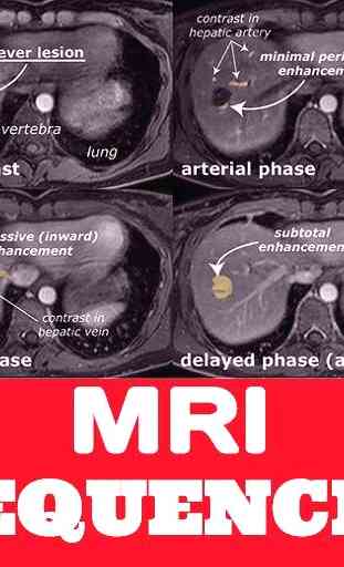
Magnetic Resonance Imaging Sequences - GUIDE
An MRI sequence is a number of radiofrequency pulses and gradients that result in a set of images with a particular appearance. This article presents a simplified approach to recognizing common MRI sequences, but does not concern itself with the particulars of each sequence.
Overview
The simplest way to think about the multitude of sequences available on modern scanners is to divide them according to the dominant influence on the appearance of tissues. This leads to a division of all sequences into proton density (PD) weighted, T1 weighted, T2 weighted, diffusion weighted, flow sensitive and 'miscellaneous'. A number of 'optional add-ons' can also be considered, such as fat or fluid attenuation, or contrast enhancement.
This leads to a broad categorisation as follows:
T1
gadolinium enhanced
fat suppressed
T2
fat suppressed
fluid attenuated
susceptibility sensitive
proton density
fat suppressed
diffusion weighted
flow sensitive
MR angiography
MR venography
CSF flow studies
miscellaneous
MR cholangiopancreatography (MRCP)
a special T2-weighted sequence
MR spectroscopy
MR perfusion
functional MRI
tractography
Terminology
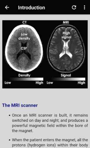
Intensity
When describing most MRI sequences we refer to the shade of grey of tissues or fluid with the word intensity, leading to the following absolute terms:
high signal intensity = white
intermediate signal intensity = grey
low signal intensity = black
Often we refer to the appearance by relative terms:
hyperintense = brighter than the thing we are comparing it to
isointense = same brightness as the thing we are comparing it to
hypointense = darker than the thing we are comparing it to
Annoyingly these relative terms are used without reference to the tissue being used as the comparison. In some instances this does not lead to any problems; for example, a hyperintense lesion in the middle of the liver is clearly hyperintense compared to the surrounding liver parenchyma. In many other situations however use of relative terms leads to potential confusion. Imagine a lesion within the ventricles of the brain described as "hypointense". Does this denote a lesion darker than CSF or than the adjacent brain?
As such it is preferable to either use absolute terminology or, if using relative terms, to acknowledge the comparison tissue e.g. "the lesion is hyperintense to the adjacent spleen".
NB: the word density is for CT, and there are few better ways to show yourself as an MRI noob than by making this mistake.
Overview
The simplest way to think about the multitude of sequences available on modern scanners is to divide them according to the dominant influence on the appearance of tissues. This leads to a division of all sequences into proton density (PD) weighted, T1 weighted, T2 weighted, diffusion weighted, flow sensitive and 'miscellaneous'. A number of 'optional add-ons' can also be considered, such as fat or fluid attenuation, or contrast enhancement.
This leads to a broad categorisation as follows:
T1
gadolinium enhanced
fat suppressed
T2
fat suppressed
fluid attenuated
susceptibility sensitive
proton density
fat suppressed
diffusion weighted
flow sensitive
MR angiography
MR venography
CSF flow studies
miscellaneous
MR cholangiopancreatography (MRCP)
a special T2-weighted sequence
MR spectroscopy
MR perfusion
functional MRI
tractography
Terminology
Intensity
When describing most MRI sequences we refer to the shade of grey of tissues or fluid with the word intensity, leading to the following absolute terms:
high signal intensity = white
intermediate signal intensity = grey
low signal intensity = black
Often we refer to the appearance by relative terms:
hyperintense = brighter than the thing we are comparing it to
isointense = same brightness as the thing we are comparing it to
hypointense = darker than the thing we are comparing it to
Annoyingly these relative terms are used without reference to the tissue being used as the comparison. In some instances this does not lead to any problems; for example, a hyperintense lesion in the middle of the liver is clearly hyperintense compared to the surrounding liver parenchyma. In many other situations however use of relative terms leads to potential confusion. Imagine a lesion within the ventricles of the brain described as "hypointense". Does this denote a lesion darker than CSF or than the adjacent brain?
As such it is preferable to either use absolute terminology or, if using relative terms, to acknowledge the comparison tissue e.g. "the lesion is hyperintense to the adjacent spleen".
NB: the word density is for CT, and there are few better ways to show yourself as an MRI noob than by making this mistake.
Category : Medical
Related searches