
MRI - Regional Anatomy of the Brain Using MRI
Magnetic resonance imaging ( MRI ) is a medical imaging technique used in radiology to form pictures of the anatomy and the physiological processes of the body. MRI scanners use strong magnetic fields, magnetic field gradients, and radio waves to generate images of the organs in the body.
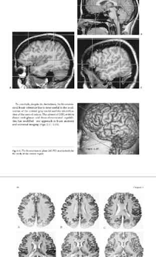
The App provides a unique review of the essential topographical anatomy of the brain from an MRI perspective, correlating high-quality anatomaical plates with the corresponding high-resolution MRI images. The App includes a historical review of brain mapping and an analysis of the essential reference planes used for the study of the human brain. Subsequent chapters provide a detailed review of the sulcal and the gyral anatomy of the human cortex, guiding the reader through an interpretation of the individual brain atlas provided by high-resolution MRI. The relationship between brain structure and function is approached in a topograhical fashion with analysis of the necessary imaging methodology and displayed anatomy. The central, perisylvian, mesial temporal and occipital areas receive special attention. Imaging of the core brain structures is included. An extensive coronal atlas concludes the app. Neuroscientists, neuroradiologists, neurologists, neurosurgeons and students of human behavior should find this app useful guiding them to a better understanding of the localization of brain function.
Brain MRI provides concise, easily accessible information on MRI physics and is an invaluable revision aid. All topics are included from magnetism to safety, K space to pulse sequences, image contrast to artefacts. The second edition has been fully revised and updated with brand new information on data acquisition and pulse sequences. The app is now in full colour throughout and follows the familiar, easy-to-use at a Glance format with each topic presented as a double-page spread with key facts accompanied by clear diagrams encapsulating essential knowledge.
Historical Review of Cross-Sectional Anatomy of the Brain
Cephalic Reference Lines Suitable for Neuroimaging
Brain Cortical Mantle and White Matter Core
Central Region and Motor Cortex
Perisylvian Cognitive Region
Limbic Lobe and Mesial Temporal Region
The Basal Forebrain, Diencephalon and Basal Ganglia
The Brainstem and Cerebellum
Optic Pathway and Striate Cortex
Atlas of Cross-Sectional Anatomy of the Brain
the app has become the standard text for radiographers, technologists, radiology residents, radiologists and even sales representatives on the subject of magnetic resonance imaging. This text is essential reading on postgraduate courses. Furthermore MRI in Practice has come to be known as the number one reference app and study guide in the areas of MR instrumentation, principles, pulse sequences, image acquisition, and imaging parameters for the advanced level examination for MRI offered by the American Registry for Radiologic Technologists (ARRT) in the USA.
The app explains in clear terms the theory that underpins magnetic resonance so that the capabilities and operation of MRI systems can be fully appreciated and maximized. This third edition captures recent advances, and coverage includes: parallel imaging techniques and new sequences such as balanced gradient echo.
Category : Education
Related searches
Reviews (3)
Sk. S. T.
Dec 6, 2021
No more information
Gop. N.
Jan 17, 2021
Highly Informative
The recent upgrade took away the ability to increase the size of the text.