NeuroNavigator
Developed by Imeka: www.imeka.ca - the only company dedicated to white matter imaging and diffusion MRI. We've been developing tools since 2011 to look at white matter microstructure and connectivity to help cure brain disease.
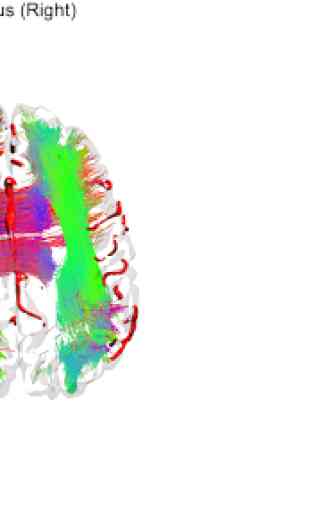
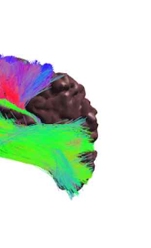
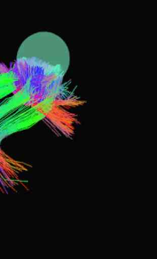
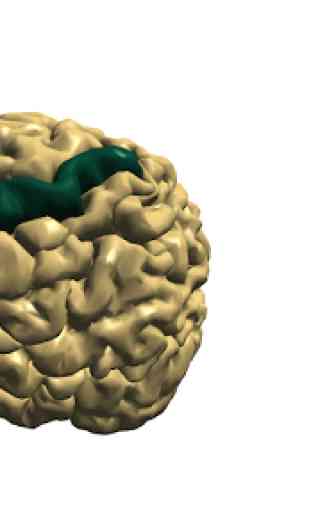
Visualize and interact with human brain white matter, gray matter, and vascular anatomy derived from cutting edge multi-modal human neuroimaging magnetic resonance imaging (MRI) datasets.
The app contains neuroimaging data from a 33 year old healthy human male brain including:
- 84 cortical areas based on gray matter segmentation of anatomical T1-weighted MRI image (T1)
- 24 subcortical areas, including ventricles and cerebellum segmented from anatomical T1
- 33 white matter pathways reconstructed using tractography from diffusion weighted MRI image (DWI)
- venous segmentation based on susceptibility weighted MRI image (SWI)
- arterial segmentation based on time of flight MRI image (TOF)
- functional connectivity based on blood oxygen level dependent (BOLD) functional MRI images
Cortical and subcortical regions of interest (ROIs) were segmented automatically using the Freesurfer software package:
https://www.sciencedirect.com/science/article/pii/S1053811912000389
White matter pathways were processed and automatically segmented using in-house software developed at Imeka, based on the following publication:
https://www.sciencedirect.com/science/article/pii/S1053811917305839
Venous and arterial segmentations were segmented automatically using in-house software developed at the University of Sherbrooke, based on the following publication:
https://onlinelibrary.wiley.com/doi/abs/10.1002/hbm.24337
Inspired by the following work:
3D interactive tractography-informed resting-state fMRI connectivity
https://www.frontiersin.org/articles/10.3389/fnins.2015.00275/full
Real-time multi-peak tractography for instantaneous connectivity display
https://www.frontiersin.org/articles/10.3389/fninf.2014.00059/full
All data was acquired with informed consent on a 3 Tesla Philips Ingenia MRI scanner at the Centre Hospitalaire Universite de Sherbrooke in Sherbrooke, Quebec, Canada.
Useful for learning about the anatomy of the brain.
Category : Education
Related searches
Reviews (8)
Ama. L.
Jun 17, 2019
Brilliant brain visualization app!! Very optimal representation of the brain anatomy, vasculature and tractography!
Ale. J.
Oct 31, 2019
Perfect app, it takes time to get use to controls but after couple minutes you realise it is what you always want.
Fél. C. M.
Feb 21, 2019
Very nice app! Smooth rendering. Very useful for education purposes. Lots of options!!!!
Phi. D.
Feb 21, 2019
Impressive images, good performance, lots of features.
Pie. J.
Feb 22, 2019
Super app to navigate within the brain in realtime. Just awesome.
Mic. B.
Feb 21, 2019
That is some amazing work, smooth and intuitive!
Chr. C.
Mar 2, 2019
Wow great app !
excellent. the best brain anatomy visualizer on the app store...very interactive with smooth graphics and a wide range of structures to choose from